Digital Dental Technology at The Dental Design Center Pattaya
▶ Digital Workflow for Implant Treatment Planning
Implant therapy has become an essential component of dental treatment for a vast number of patients. One change that reshaped the clinical approach to implant treatment planning and therapy was the introduction of digital technologies and workflows. Due to their advantages in accurate diagnosis, treatment planning, and executing implant procedures, digital technologies are becoming more widely utilized. Elements of a digital workflow include data collection, computer aided design/computer aided manufacturing , and implant placement .These digital advancements have gained widespread adoption due to their numerous advantages in precise diagnosis, treatment planning, and the execution of implant procedures.
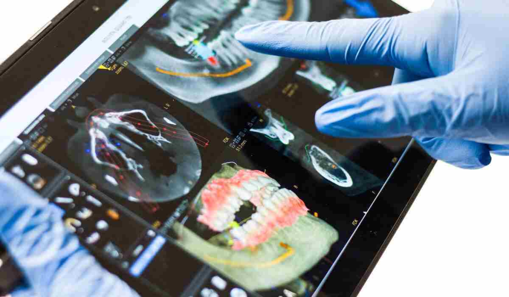
A digital workflow for implant treatment planning is a streamlined and precise process that leverages advanced digital technologies to plan and execute dental implant procedures. This approach offers numerous benefits, including enhanced accuracy, efficiency, and patient comfort. Here’s an overview of the digital workflow for implant treatment planning:
Digital Imaging: The process typically begins with the acquisition of high-resolution digital images. This may include cone-beam computed tomography (CBCT) scans, intraoral scans, and extraoral scans, depending on the patient’s specific needs. These digital images provide detailed information about the patient’s oral anatomy, bone structure, and tooth positioning.
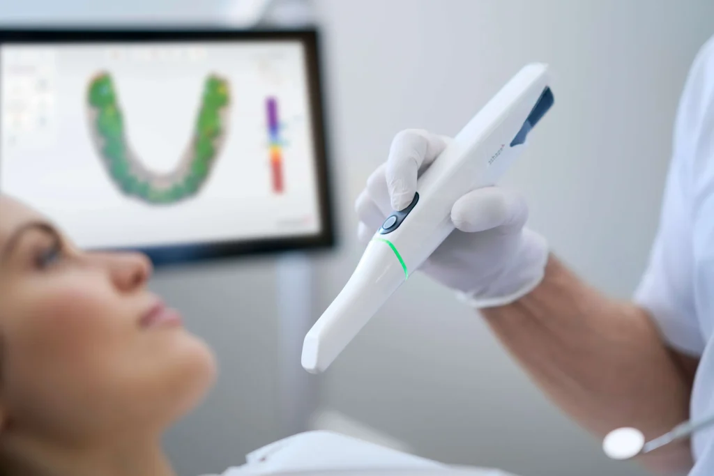
Digital Impressions: Intraoral scanners are used to create digital impressions of the patient’s teeth and gums. This eliminates the need for traditional messy impression materials and offers greater comfort to the patient.
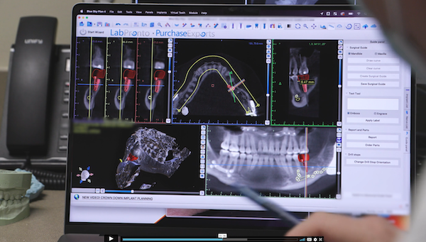
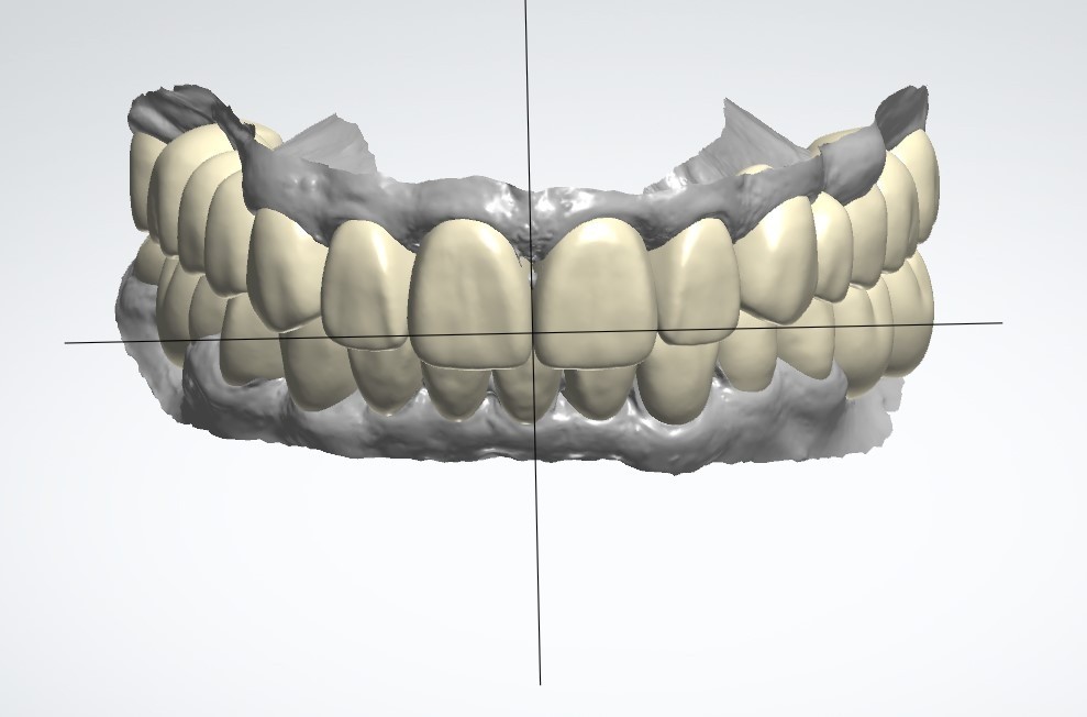
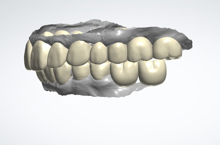
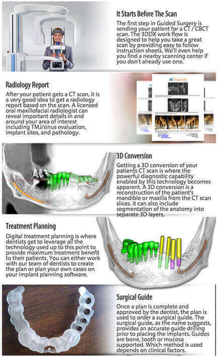
Software-Based Planning: Specialized software is employed to process the digital data and create a 3D model of the patient’s oral anatomy. This 3D model allows the dental team to precisely plan the implant placement, taking into account factors such as bone density, available space, and aesthetics.
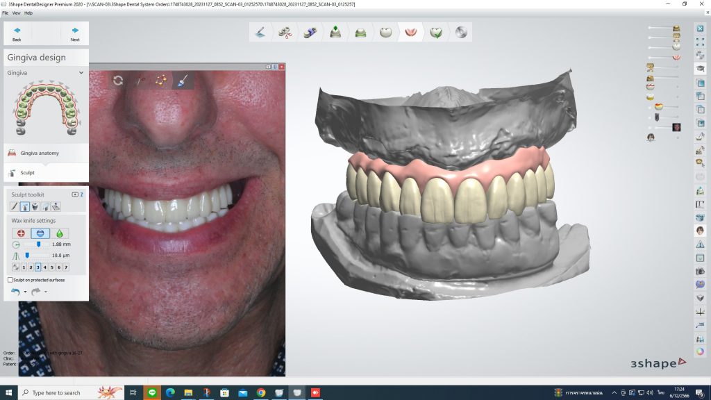
Virtual Implant Placement: Using the 3D model, the dentist can virtually place the dental implants in the ideal positions. They can adjust the angle, depth, and orientation of the implants for optimal stability and function. This tool facilitate our dental implant specialist in All-on-4 implants procedure planning. .
Digital Surgical Guides: Digital planning software can generate surgical guides, which are custom-made templates that guide the dentist during the implant surgery. These guides ensure that the implants are placed exactly as planned, minimizing the margin for error.
Patient Communication: Digital technology allows for clear and visual communication with the patient. Dentists can show patients the 3D model, explain the treatment plan, and help them understand what to expect from the procedure.
Fabrication of Prosthetics: After the implants are placed, digital impressions can be used to design and fabricate custom prosthetics, such as crowns, bridges, or dentures. This ensures a precise fit and natural appearance.
Follow-Up and Monitoring: Digital records of the implant procedure are stored electronically, allowing for easy access and monitoring during follow-up appointments.
Collaboration: Digital workflows also facilitate collaboration between different specialists involved in the implant procedure, such as oral surgeons, prosthodontists, and dental laboratory technicians. They can share digital files and collaborate more effectively.
Patient Records: All digital data, including images, treatment plans, and patient records, can be securely stored in a digital format, simplifying record-keeping and ensuring data integrity.
Overall, a digital workflow for implant treatment planning enhances precision, reduces treatment time, and improves the overall patient experience when compared to traditional analog methods. It has become a standard practice in modern implant dentistry.
The advent of digital workflows for implant treatment has increased accuracy and improved diagnosis, treatment planning and surgical execution in dental implant placement. In turn, these benefits have led to more predictable and successful implant outcomes.
Digital Intraoral Scanner
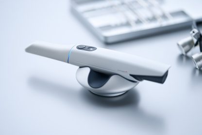
The newest digital intraoral scanner at The Dental Design Clinic Pattaya: TRIOS 5 from 3 Shape (Denmark) TRIOS 5 This is the scanner that sets a new standard in patient protection and infection control. It is not only hygienic by design, but also smaller, lighter, and designed to fit perfectly in every hand. On top of that, it comes with our new ScanAssist engine for perfect scan results, and TRIOS Share, for ultimate connectivity. Simply hygienic. Simply ergonomic.
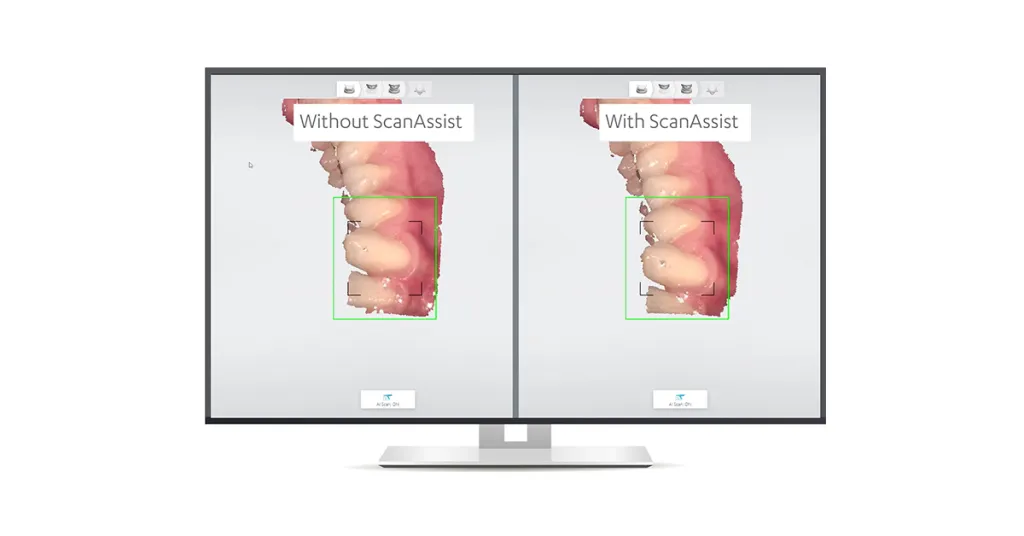
Key features of the 3Shape TRIOS 5 intraoral scanner may include:
- High-Quality Scanning: The scanner captures precise digital impressions quickly and efficiently, allowing dental professionals to create accurate treatment plans.
- Color Scanning: which can help with shade matching and cosmetic treatments.
- Real-Time Feedback: Dentists can review scans in real-time, ensuring that they have captured all necessary data before completing the procedure.
- Digital Workflow: The digital impressions can be seamlessly integrated into CAD/CAM systems for designing restorations like crowns, bridges, and orthodontic appliances.
- Patient Engagement: The 3Shape TRIOS system often includes patient-friendly features, such as an interactive touchscreen, which can improve patient engagement and comfort during the scanning process.
- Compatibility: The scanner is typically compatible with various dental software programs and systems, making it versatile for different clinical needs.
- The 3Shape TRIOS intraoral scanners have become popular in modern dentistry due to their accuracy, efficiency, and the ability to enhance the overall quality of dental care. They have significantly improved the precision and convenience of digital impressions, benefiting both dental professionals and patients.
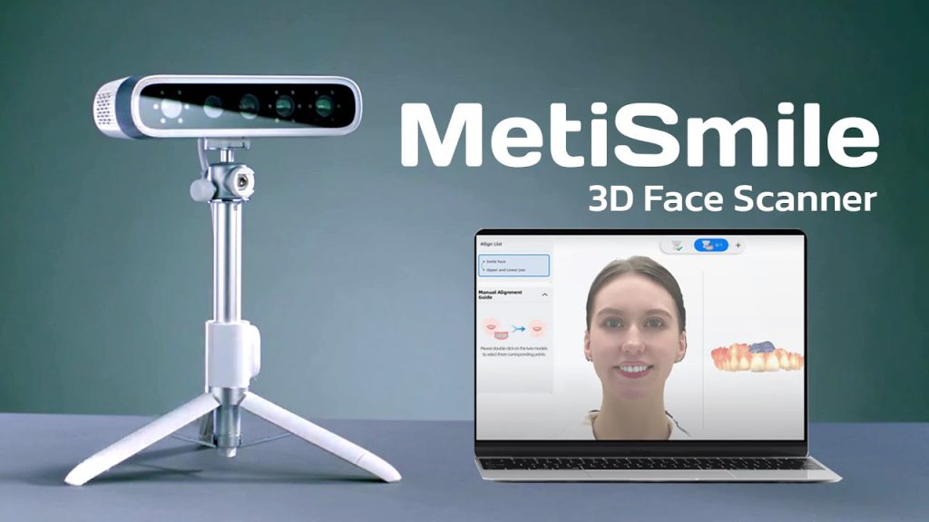
Dental Digital Face Scan
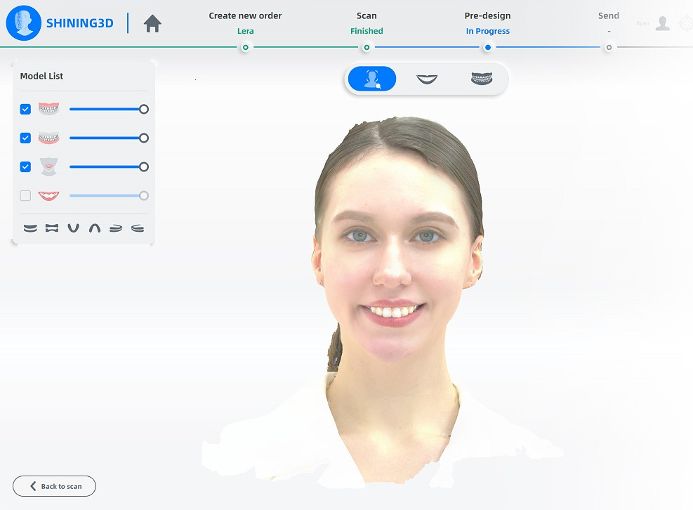
A dental digital face scan is a modern technique used in dentistry to capture precise three-dimensional images of a patient’s face and oral structures. This process typically involves using specialized equipment, such as intraoral scanners or facial scanners, to create highly detailed digital models. These scans are invaluable in various dental procedures, including treatment planning for orthodontics, dental implants, and cosmetic dentistry.
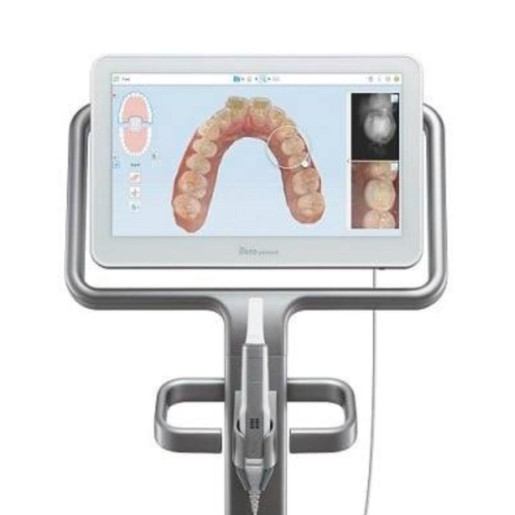
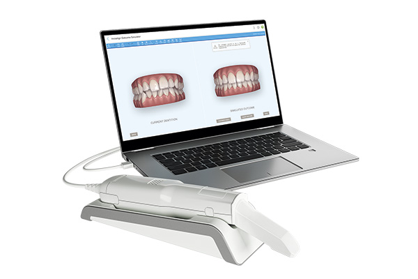
iTero Element™ 5D
iTero Element™ 5D imaging system benefits.
One imaging system does it all. 3D digital impressions. Crisp intraoral scanning. Treatment simulation. And more – without harmful radiation.
Advanced technology to scan internal tooth structure. The first 3D intraoral scanner with Near-Infrared Imaging (NIRI) technology to aid your diagnostic workflow. Aids in detection and monitoring of interproximal caries above the gingiva in real time. Show your patients how their treatment is progressing with iTero TimeLapse technology. When patients can see their changes over time, they’re more likely to stay engaged and take better care of their oral health. Which helps you to keep an eye on treatment progress, for better outcomes.
▶ CT Technology at the Dental Design Center
A CT (computed tomography) scan, also called a CAT (computerized axial tomography) scan, is a diagnostic aid used to obtain three-dimensional images of various body structures. It is a specialized kind of X-ray which is of high diagnostic value because of its clarity and accuracy. Many dental professionals use this type of scan in conjunction with diagnosing and treating their patients.
▶ Anthos ergonomic dental chair
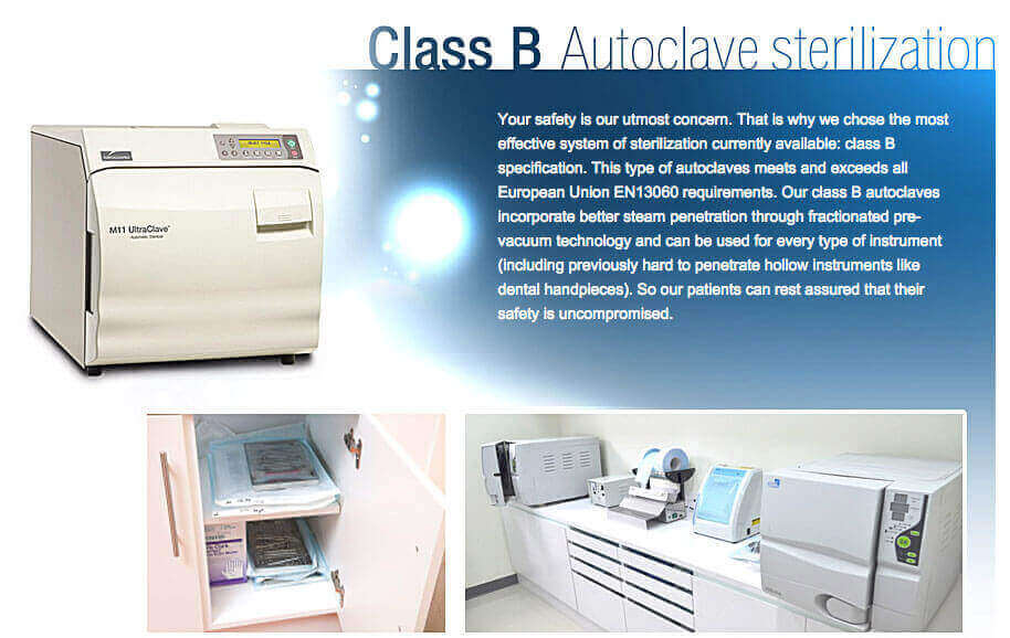
Our sterilized instruments are stored in purpose-designed storage cabinets that can be easily cleaned Before being used, our instruments are checked to ensure that:
- the packaging is intact
- the sterilization indicator confirms the pack has been subjected to an appropriate sterilization process
As part of our essential sterilization quality requirements, instruments that have remained unused for more than 21 days and are not in a validated sterile pack (processed by a vacuum sterilizer) should be subjected to a further cycle of decontamination before being used.
▶ Dental Implant Armamentarium
Piezotome
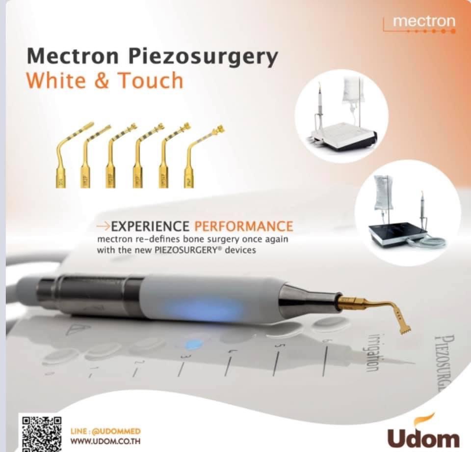
Piezosurgical is a process that utilizes piezoelectric vibrations in the application of cutting bone tissue. By adjusting the ultra sonic frequency of the device, it is possible to cut hard tissue while safely avoiding damage to nearby soft tissues. This decrease in collateral trauma during procedures such as osteotomies, osteoplasties, ridge expansion, autogenous bone harvesting, and sinus lifts results in rapid healing and a more comfortable and predictable recovery for our patients.
The innovative qualities of the PIEZOSURGERY
- Micrometric cutting action for maximum surgical precision and intra-operative sensitivity.
- Selective cutting action for minimal damage to soft tissue, maximum safety for you and your patients.
- Cavitation effect maximum intra-operative visibility and a blood-free surgical site.
This innovative dental technology is very useful in dental implant surgery for example sinus lifting for dental implant placement, tooth extraction, bone splitting and bone grafting procedure, etc.
▶ Osstell
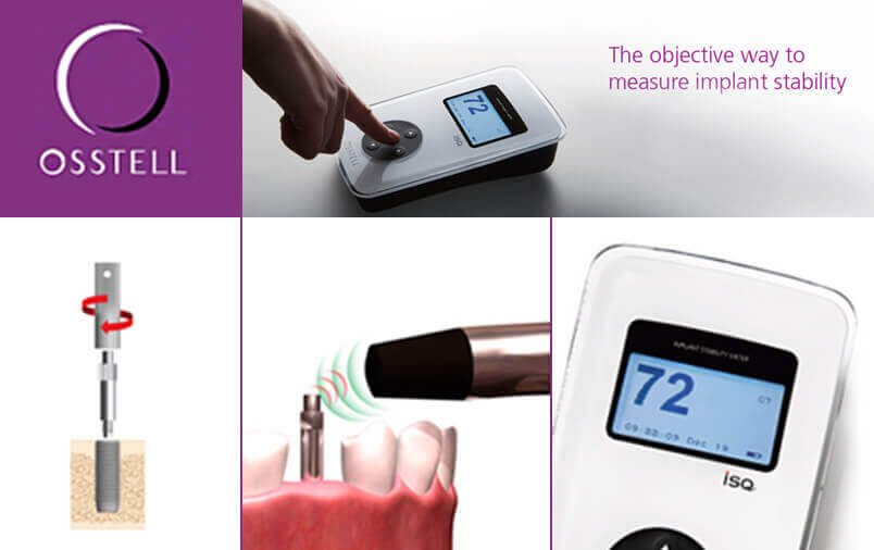
The degree of implant stability is hard to determine once the implant has started to integrate. Also, at placement, stability is difficult to judge objectively and the “tactile feeling” or torque measurements will not work as a baseline for future comparisons. Osstell meters make it possible not only to measure the initial implant stability, but also to monitor the development of osseointegration over time.
Benex: Atraumatic tooth extraction system
▶ PRF Centrifuge
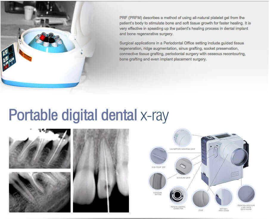
Portable digital dental x-ray improves dental radiography speed, convenience and quality. The Dexcowin DX-3000 technology provides the safest and highest quality handheld intraoral X-ray system.
An x-ray can be taken at anytime during dental procedures for verification, particularly in endodontic treatment and dental implant surgery. Dentists will have more diagnostic information for a continuous work. With the Dexcowin DX-3000 we can bring the x-ray to the patient, not the patient to the x-ray – The patient doesn’t even leave the dentist chair! Saves time and improved workflow!
▶ NiTi rotary files: ProTaper (Dentsply-Maillefer) systems
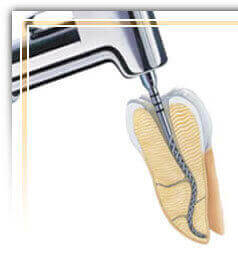
Niti alloy files have quite simply revolutionised our ability to predictably clean and shape the root canal system. In an electric (torque-controlled) motor and endodontic handpiece.
▶ Apex locators
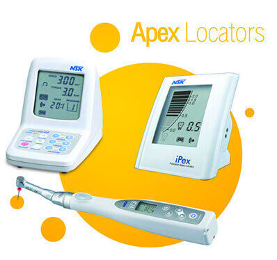
Apex locators have come along way in the last 10 years and now give a very predictable ‘linear view’ of a 3-dimensional root canal. We know that the apical constriction of a root is never at the radiographic apex, hence the dictum of working the average “1 mm away from the radiographic apex”. This usually keeps us out of trouble. But, for absolute certainty the Root ZX II tells us exactly when we have reached the hallowed apical constriction in 90-95% of cases..
▶ Sedation Dentistry for Dental Treatments

The Dental Design Center offers mild sedation dentistry (nitrous oxide or laughing gas sedation) to reduce dental anxiety and dental fear during treatment.
DDC, we are committed in provide patients with safe and effective use of anesthesia for investigation, procedure and dental treatment (Local anesthesia, mild sedation).
Our dentists comply with standard and professional qualifications.
Proudly powered by WordPress
